


Autism and Brain Scans: What Neuroimaging Reveals About the Spectrum
Autism Spectrum Disorder (ASD) affects how people communicate, socialize, and process the world around them. While diagnosis often relies on behavioral observations, autism and brain scans are opening new doors to understanding the condition at a neurological level. Advanced imaging like MRI and fMRI shows distinct brain differences in autistic individuals, from enlarged brain volumes in toddlers to altered connectivity patterns. This article explores how brain scans illuminate autism, their diagnostic potential, and what they mean for therapies like ABA and occupational therapy.
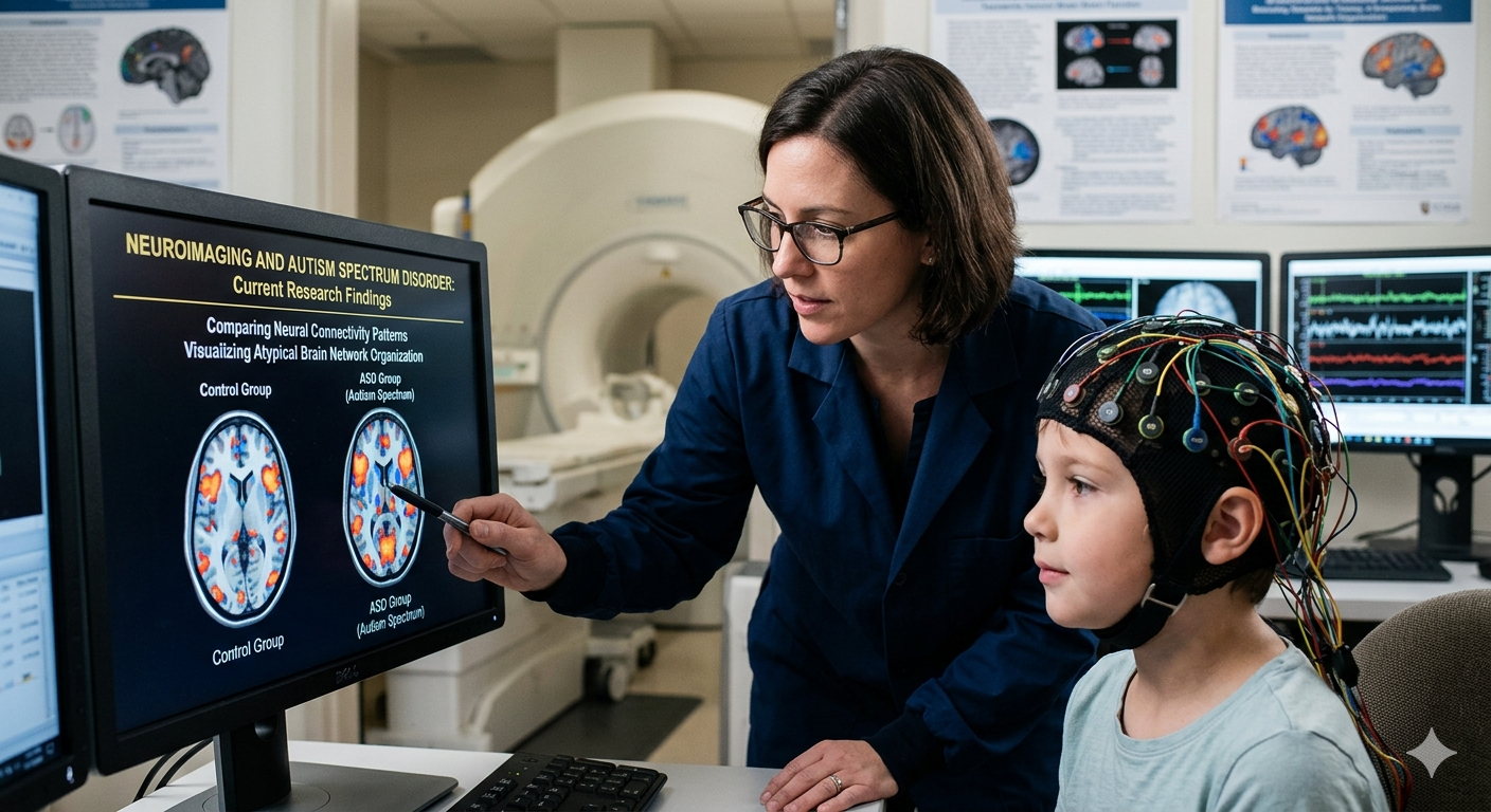
The Science Behind Brain Scans for Autism
Brain scans use non-invasive techniques to peer inside the living brain, revealing structures and activities invisible to the naked eye. For autism research, common tools include:
-
Structural MRI: Measures brain anatomy, spotting differences in size, shape, and white matter.
-
Functional MRI (fMRI): Tracks blood flow to active brain regions during tasks, highlighting social and sensory processing.
-
Diffusion Tensor Imaging (DTI): Maps white matter tracts, showing how brain areas “talk” to each other.
Studies, such as those from the Infant Brain Imaging Study (IBIS), have found that infants later diagnosed with autism often show rapid brain overgrowth by age 2-3. For instance, a 2023 meta-analysis in JAMA Pediatrics reviewed over 100,000 scans and confirmed enlarged cortical surfaces in 80% of high-risk toddlers with ASD.
These findings challenge the idea of autism as purely behavioral—they point to tangible brain differences emerging early in life.
Key Brain Differences Uncovered by Scans
Autism and brain scans consistently reveal patterns across the spectrum:
-
Brain Overgrowth: Toddlers with autism have 10-15% larger brain volumes, especially in the frontal and temporal lobes, per NIH-funded research. This “head growth spurt” peaks around 12 months.
-
Connectivity Issues: DTI scans show “underconnectivity” in long-range tracts (e.g., between hemispheres) but “overconnectivity” locally. A landmark 2024 study in Nature Neuroscience linked this to sensory overload and social challenges.
-
Amygdala and Social Processing: The amygdala, key for emotions, is often enlarged early but shrinks by adolescence, correlating with anxiety in ASD, as seen in fMRI studies from UCLA.
-
Default Mode Network (DMN) Disruptions: fMRI reveals weaker DMN activity during rest, explaining mind-wandering differences and repetitive behaviors.
These aren’t universal—autism’s heterogeneity means scans vary by individual, sex, and severity.
Can Brain Scans Diagnose Autism?
Not yet reliably for everyday use. The FDA hasn’t approved scans as standalone diagnostics; they complement tools like the ADOS-2. However:
-
Early Detection: A 2025 AI model from Boston Children’s Hospital analyzed 1,000+ infant MRIs with 95% accuracy in predicting ASD at 6 months—months before behavioral signs.
-
Subtyping: Scans help classify “high-functioning” vs. severe cases, guiding personalized interventions.
-
Ruling Out Mimics: They distinguish autism from epilepsy or genetic syndromes like Fragile X.
Challenges remain: High costs ($1,000-$5,000 per scan), need for sedation in young kids, and variability across scanners. Ongoing trials, like EU-AIMS, aim for biomarkers by 2030.
Therapeutic Insights from Autism Brain Scans
Brain imaging doesn’t just diagnose—it informs treatment. Therapists use scan data to tailor approaches:
-
ABA Therapy: fMRI shows ABA strengthens DMN connectivity, improving social reciprocity.
-
Occupational Therapy: Scans reveal sensory integration issues in parietal lobes, so OTs target tactile desensitization.
-
Speech Therapy: Reduced Broca’s area activation guides language interventions with visual supports.
-
Emerging Tech: Neurofeedback uses real-time fMRI to train self-regulation, with a 2024 Autism Research pilot showing 30% symptom reduction.
For example, a child with overactive amygdala on scans might benefit from mindfulness apps calibrated to fMRI patterns.
Limitations and Ethical Considerations
Autism and brain scans hold promise, but beware hype. Scans capture averages, not individuals—neurodiversity advocates argue they pathologize natural variation. Privacy risks loom with AI databases, and access disparities hit underserved communities hardest.
Experts like Dr. Catherine Lord emphasize: “Scans are tools, not labels.” Combine them with behavioral expertise for holistic care.
The Future of Neuroimaging in Autism
With AI advancing, expect portable EEG-fMRI hybrids and prenatal risk prediction. Projects like the BRAIN Initiative fund scans linking genetics (e.g., SHANK3 mutations) to brain changes. By 2030, scans could enable precision medicine, matching therapies to brain profiles.
In summary, autism and brain scans demystify a complex disorder, proving it’s rooted in biology. For parents or therapists, they offer hope: early insights lead to better outcomes.
Sources: NIH, Autism Speaks, The Lancet Neurology* (2024 reviews). Consult a specialist for personalized advice.*